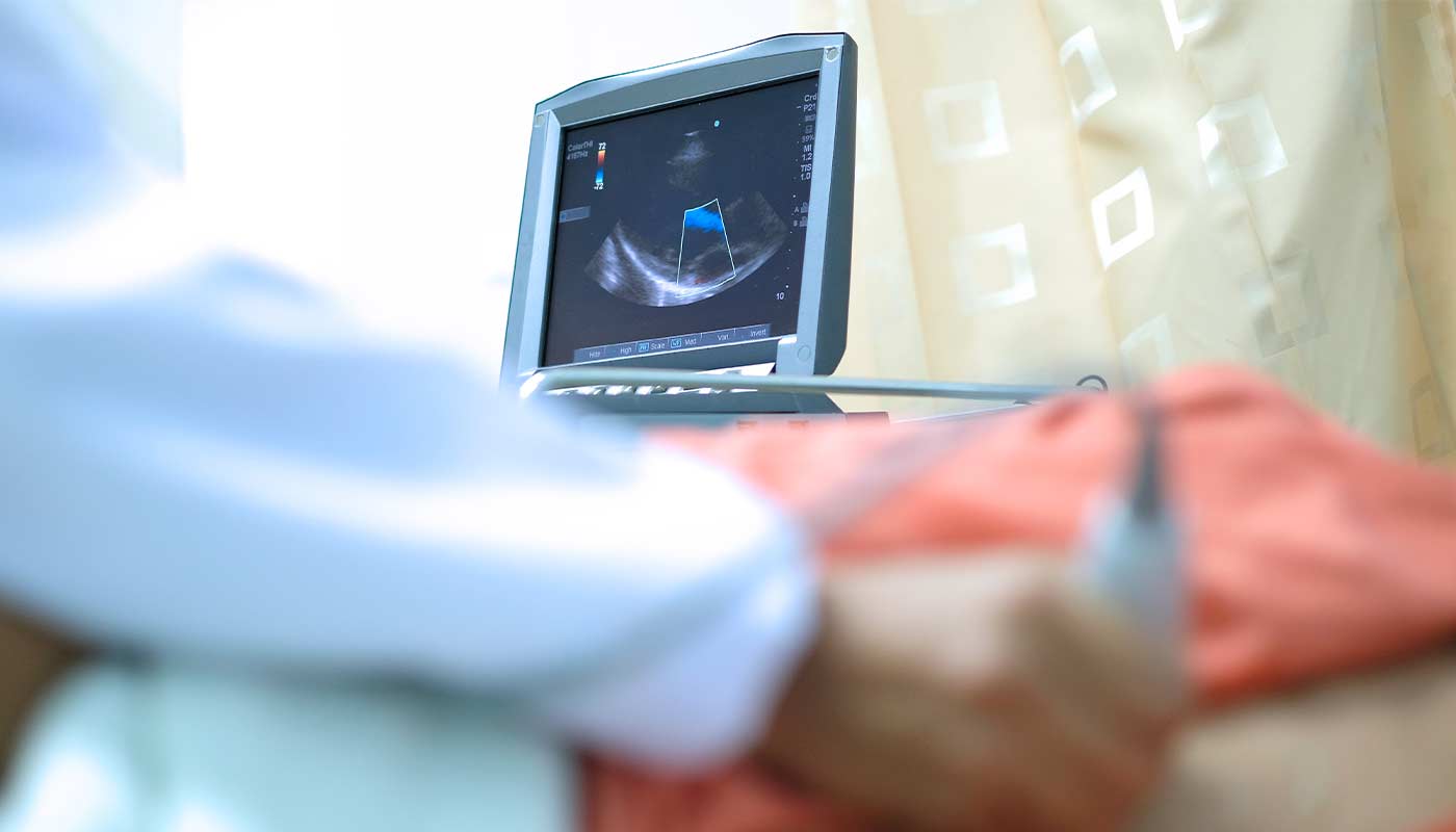
An echocardiogram is a test that uses ultrasound waves to create images of the heart. This examination helps assess the structure and function of the heart. Continue reading to learn more about heart echocardiograms.
Reasons for Performing Echo
Doctors may recommend a heart echo for various reasons, such as:
- Diagnosing the cause of symptoms that may be linked to heart disorders.
- Diagnosing heart disease.
- Monitoring the condition of patients with heart diseases, such as valve disorders that require regular follow-up.
- Assessing heart function before undergoing surgery.
- Evaluating the results of surgeries or medical procedures related to the heart.


What Does an Echo Reveal?
- Congenital heart defects.
- Issues related to the heart’s pumping function or its ability to relax between beats.
- Cardiomyopathy.
- Infective endocarditis.
- Pericarditis (inflammation of the sac surrounding the heart).
- Heart valve diseases.
- Detecting any heart changes that might indicate aortic aneurysms, blood clots, or heart tumors.
Types of Echo
There are several types of echoes, each used based on the patient’s condition and the test’s purpose. Below are some types and the steps for each:
- Transthoracic Echo (TTE): This is the most common and widely used type. During this exam, a handheld device called a transducer is used to send ultrasound waves to the patient’s chest, capturing several images of the heart. No specific preparations, such as fasting or medication adjustments, are required for this test unless instructed by your doctor.
- Transesophageal Echo (TEE): This type is used when more detailed and accurate images are needed. A transducer is inserted through the esophagus to get clearer heart images, especially if there are difficulties in imaging through the chest. Before the test, you should inform your doctor if you have sleep apnea, swallowing problems, esophageal issues such as a hiatal hernia, or if you’re taking medications for anxiety, sleep disorders, or pain relief. Avoid eating or drinking for at least 6 hours before the test.
- Stress Echo: A stress echo is performed before and after exercise in a hospital or clinic to assess how the heart responds to physical activity. If the patient is unable to exercise, medication is administered to make the heart work harder, simulating the effects of exercise.
Here are some guidelines to follow before the test:
- Avoid eating or drinking, except for water, for at least 4 hours before the test.
- Avoid smoking on the day of the test.
- Avoid caffeine for 24 hours before the test, including decaffeinated drinks and some over-the-counter pain relievers.
- Inform your doctor about all medications you’re taking; they may recommend stopping certain heart medications and adjusting diabetes medication doses on the day of the test.
- Wear comfortable clothing and athletic shoes.
Echo Results
The results of an echocardiogram may indicate various heart problems, such as valve issues, heart muscle damage, blood clots in the heart, or problems with blood flow during exercise. If any of these issues are detected, the doctor may recommend further tests before diagnosing a heart disorder or creating a treatment plan if a heart disorder is diagnosed.
In conclusion, echocardiograms are essential medical tests that help diagnose many heart diseases safely and effectively. Through this test, doctors can obtain a clear and comprehensive picture of heart function, contributing to appropriate treatment plans to maintain heart health.
Resourses
https://my.clevelandclinic.org/health/diagnostics/16947-echocardiogram https://www.mayoclinic.org/tests-procedures/echocardiogram/about/pac-20393856 https://www.healthline.com/health/echocardiogram
